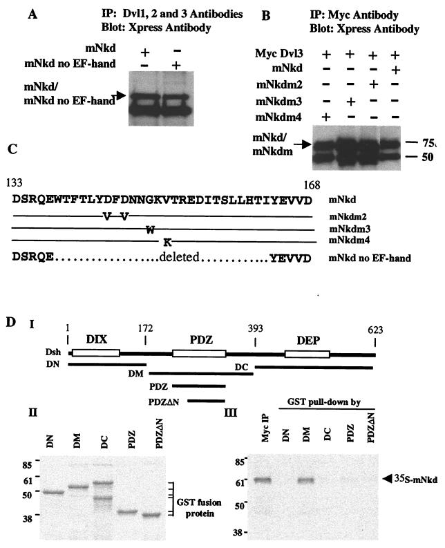
Figure 3.
mNkd interacts with Dishevelled. (A) Lysates were prepared from Cos7 cells expressing mNkd, mNkd no EF-hand, or GFP constructs. Total cell lysates were immunoprecipitated with monoclonal antibodies 1, 2, and 3 to Dishevelled (Santa Cruz Biotechnology); after SDS/PAGE and membrane transfer, the membrane was blotted with Xpress antibody (Invitrogen). (B) mNkd mutations in the EF-hand do not have a significant effect on the binding of mNkd to Dishevelled. Cell lysates prepared from HEK293 cells expressing Myc-tagged Dishevelled and Xpress-tagged mNkd proteins were immunoprecipitated with Myc antibody (Roche Molecular Biochemicals); after SDS/PAGE and membrane transfer, the membrane was blotted with Xpress antibody. (C) Summary of mutations in the EF-hand of mNkd. The mNkd mutant m2 contains amino acid changes D144V and D146V. The mNkd mutant m3 contains amino acid change G149W. The mNkd mutant m4 contains amino acid change V151K. –, identical amino acids as the wild type; … . . , deleted amino acids in the mNkd no EF-hand mutant. (D) mNkd binds to the conserved middle region of Dishevelled. (I) Schematic diagram of GST-fusion proteins containing different regions of Drosophila Dishevelled (Dsh). (III) Myc-tagged mNkd protein, labeled with 35S, was immunoprecipitated with Myc antibody or precipitated by GST-fusion proteins. The immunocomplexes were separated by SDS/PAGE and detected by autoradiography. (II) Equal amounts of GST-fusion proteins used in III were separated by SDS/PAGE and detected by Coomassie blue stain.

