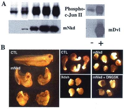
Figure 5.
mNkd activates JNK in mammalian cell culture and alters convergent extension movements in Xenopus laevis. (A) The JNK assay was carried out as described (15). NIH 3T3 cells were transfected with plasmids expressing c-Jun and mNkd or c-Jun and mDvl3. Cell lysates were separated by SDS/PAGE and transferred onto a nitrocellulose membrane. The membrane was blotted with PhosphoPlus c-Jun (Ser-63) II antibody (New England Biolabs). The same membrane was then stripped and blotted with Xpress antibody (Invitrogen) to detect the amount of Xpress-tagged mNkd or blotted with Myc antibody to detect Dishevelled. (B) Dorsal expression of mNkd in developing Xenopus embryos inhibited the normal elongation and straightening of the anteroposterior axis (Left; control embryo at top, mNkd-injected embryos below). (Center Upper) Control open-face Keller explants of the dorsal marginal zone elongated and changed shape significantly. (Right Upper) Explants expressing mNkd failed to elongate or to change shape. (Center Lower) Overexpression of wild-type Xdsh mimicked overexpression of mNkd. (Right Lower) Downstream activation of the canonical Wnt pathway by coexpression of dominant-negative GSK-3β did not attenuate the inhibitory effect of mNkd on convergent extension. Expression of dominant-negative GSK-3β alone had no effect on convergent extension (data not shown).

