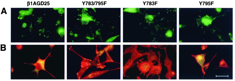
Figure 4.
Visualization of focal contacts and associated structures in v-src-transformed β1A-GD25 or mutant β1A cells on fibronectin-coated substrata. (A) Single immunofluorescent detection of β1A. (B) Double immunofluorescent detection of vinculin (green) and rhodamine-phalloidin (red). β1A-GD25, GD25 cells expressing wild-type β1A; other cells are designated by mutation(s). Microscopic analysis of multiple cells in multiple fields indicated the β1- and vinculin-containing focal contacts were present in >40% of transformed Y783F and Y783/795F cells but in <5% of β1A cells and <20% of Y795F cells. (Bar = 30 μm.)

