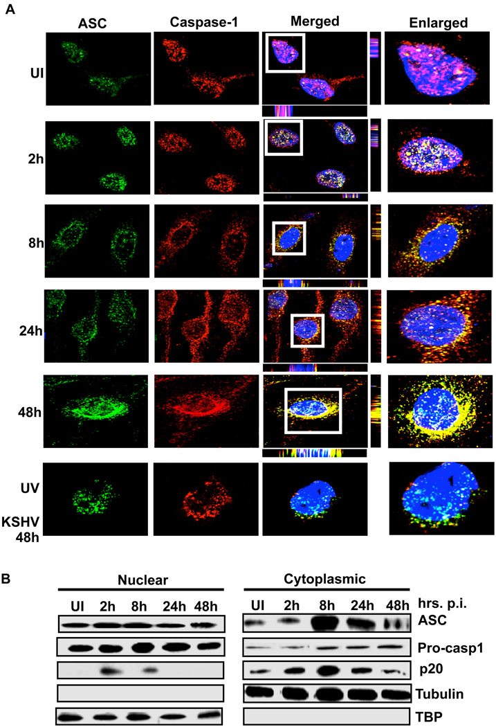
Figure 2. Inflammasome activation and sub-cellular redistribution of ASC and caspase-1.
(A) HMVEC-d cells were uninfected (UI) or infected with either live KSHV or UV-KSHV for 2 h, washed, incubated at 37°C for the indicated time, washed, fixed in 2% paraformaldehyde for 10 min, permeabilized with 0.2% Triton X-100 for 5 min and blocked with signal enhancer. Cells were reacted with anti-ASC and anti-caspase-1 antibodies, washed and incubated with Alexa-488 (green) and Alexa-594 (red) secondary antibodies, respectively. Cell nuclei were visualized by DAPI (blue). The boxed areas were enlarged and shown in the far right panel. (B) HMVEC-d cells were infected with KSHV (30 DNA copies/cell) for 2h, washed and incubated at 37°C for the indicated time. Nuclear and cytoplasmic extracts were examined by immunoblotting with anti-ASC and anti-caspase-1 antibodies. These membranes were stripped and immunoblotted with anti-tubulin and anti-TATA binding protein (TBP) antibodies to check the purity of cytoplasmic and nuclear lysates, respectively and to confirm equal loading.

