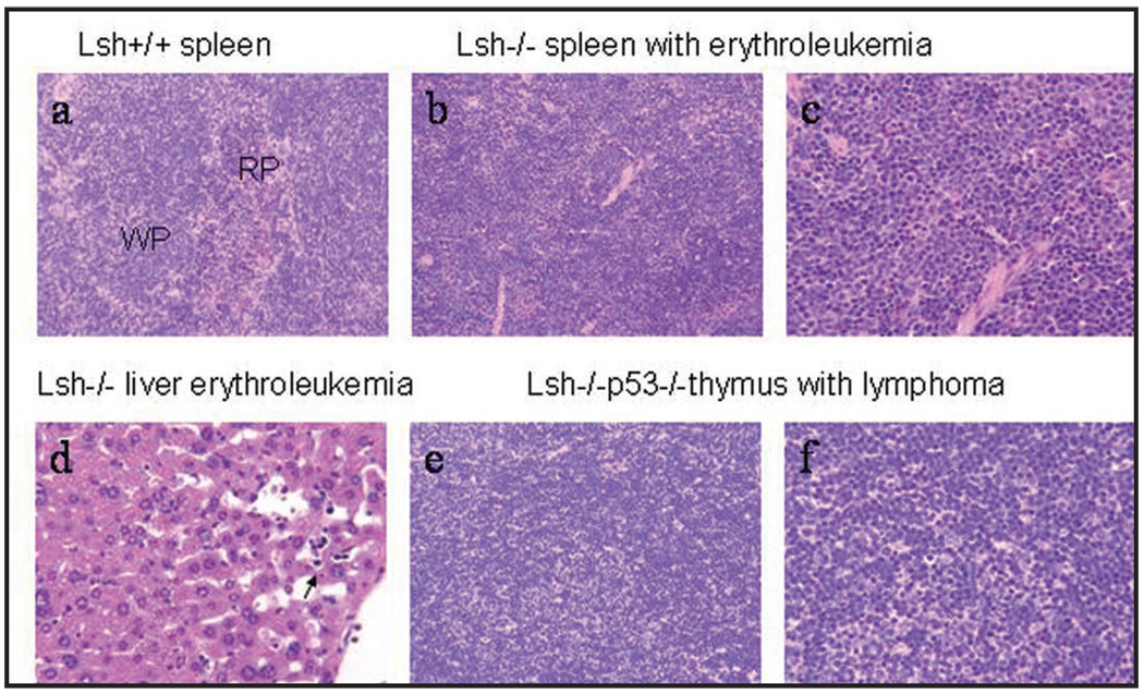
Figure 2.
Histopathological analysis of erythroleukemia and lymphoma in fetal liver cell transplanted mice. (a) Normal Spleen from mice receiving Lsh+/+ fetal liver cells. Lymphoid tissue in white pulp (wp) and normal extramedullary hematopoiesis in red pulp (rp) (H&E 20× objective). (b) Spleen with erythroleukemia from mice transplanted with Lsh−/− fetal liver cells. Effacement of white pulp by expansion of erythropoiesis originating in the red pulp. This hematopoietic neoplasm occurred in mice transplanted with Lsh−/− and Lsh−/− p53−/− fetal liver cells (H&E, 20× objective). (c) Spleen with erythroleukemia from mice transplanted with Lsh−/− fetal liver cells. Many erythrocyte precursors (cells with prominent nucleoli) as well as numerous metarubricytes (dark nuclei) (H&E, 40× objective). (d) Liver erythroleukemia from mice transplanted with Lsh−/− fetal liver cells. Circulating metarubricytes in hepatic sinusoids can be seen (arrow) (H&E, 40× objective). (e) T cell lymphoma from mice transplanted with Lsh−/−p53−/− fetal liver cells (H&E, 20×, objective). (f) Thymus with sheets of lymphoblasts from mice transplanted with Lsh−/−p53−/− fetal liver cells. (H&E, 40× objective).

