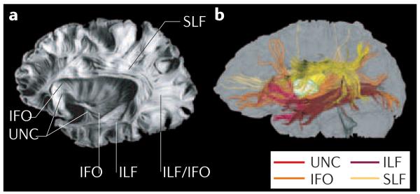
Figure 6. Comparison between a post-mortem brain sample and the results of DTI-based three-dimensional tract reconstruction.
a | Post-mortem sample showing 4 main association fibres: the superior longitudinal fasciculus (SLF), inferior longitudinal fasciculus (ILF), inferior fronto-occipital fasciculus (IFO) and uncinate fasciculus (UNC). b | These tracts can be reconstructed from in vivo human DTI data and presented with different colours. There is excellent agreement between the two. Panel a reproduced, with permission, from REF. 196 © (1997) Univ. of Iowa.

