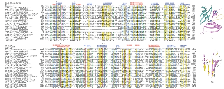
Figure 4.
(A) Multiple sequence alignment of the EndoU family emphasizing the new bacterial versions found in this study. The eukaryotic EndoU domain (PDB: 2c1w) is shown to the right to indicate the spatial position of the conserved elements and the two units with three-strands each. (B) Multiple sequence alignment of the newly identified REase family. The structure of the archaeal Holliday junction resolvase (PDB: 1OB8) is shown to the right to indicate the spatial location of the conserved residues in this fold. Secondary structure elements are indicated above the alignments (‘e’ in blue, β-sheet; ‘h’ in red, α-helix). The numbers in brackets represent excluded residues from sequences and ‘hash’ indicates the catalytic residues.

