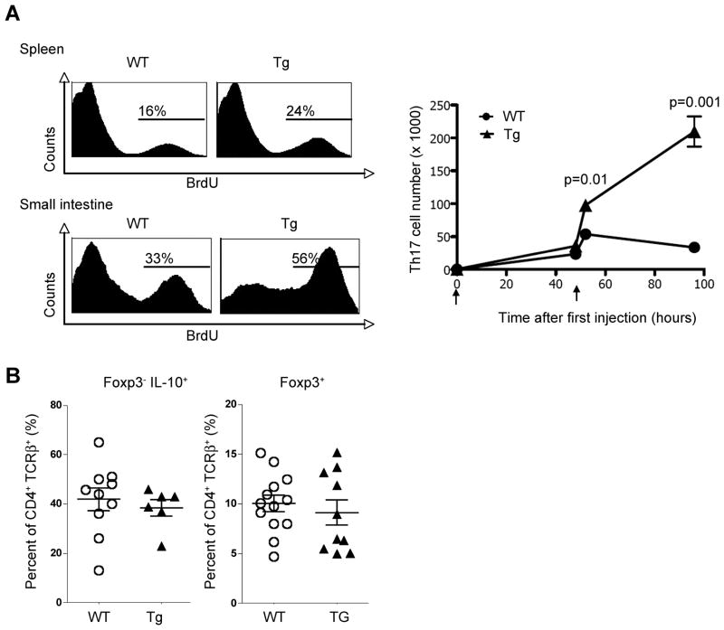
Figure 4. IL-10 signaling in T cells inhibits the proliferation of IL-17A producing cells.
(A) BrdU uptake was measured in CD4+TCRβ+IL-17A eGFP+ cells isolated from WT and Cd4-DNIL-10R mice (left panel). BrdU was injected 12 hours before mice were sacrificed. Time course experiment of total numbers of CD4+TCRβ+IL-17A+ cells after CD3-specific antibody treatment (Mean ± SEM; WT: 0h, n=4; 48h, n=2; 52h, n=5; 96h, n=5; Tg: 0h, n=4; 48h, n=2; 52h, n=4; 96h, n=5) (right panel). (B) Foxp3 RFP and IL-10 eGFP expression was measured in freshly isolated cells. Cells are gated on CD4+TCRβ+ events. A (left panel) and B: Mice were analyzed 52 hours after the first anti-CD3 injection. A (right panel) and B: Data are cumulative from three independent experiments. A (left panel): Data are representative of three independent experiments.

