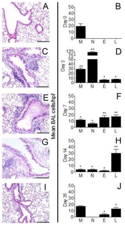
Figure 4.

Inflammation in and around the conducting airways of sensitized BALB/c mice before and after airborne conidia challenge. Lungs from sensitized BALB/c mice (A) or sensitized lungs removed at day 3 (C), 7 (E), 14 (G), or 35 (I) after a 10min inhalation dose of mature A. fumigatus conidia were stained with hematoxylin and eosin to reveal peribronchial inflammation, bar = 200 μm. Macrophage (M), neutrophil (N), eosinophil (E), or lymphocyte (L) cells washed from the airways of sensitized mice (B) or at days 335 after conidia (D,F,H,J) were cytospun onto microscope slides and differentiated by morphometric and staining characteristics. Histology and BAL cell counts for naïve mice were similar to day0 controls (not shown). Data are expressed as the mean number of cells per hpf ± SEM, n = 35 mice/group. *, p<0.05; **, p<0.01 as compared to day0 controls.
