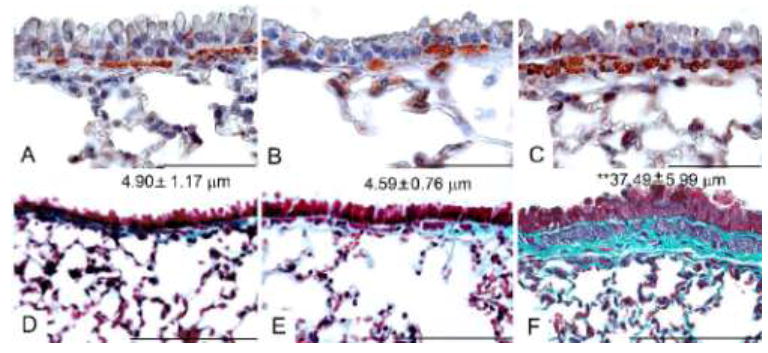
Figure 6.

Smooth muscle and fibrotic changes signifying airway wall remodeling after airborne conidia allergen challenge. αSMA staining (red) was used to identify peribronchial smooth muscle changes in histological sections from naïve BALB/c mice (A), mice sensitized to Aspergillus antigens (B), or sensitized mice that had been challenged with a 10min dose of inhaled conidia 35 days before (C). Gomori’s trichrome stain was used to show the accumulation of peribronchial collagen (blue) on histological sections from naïve mice (D), mice sensitized to Aspergillus antigens (E), or sensitized mice that had been challenged with a 10min dose of inhaled conidia 35 days before (F). Mean thickness of collagen is indicated for each group ± SEM. **, p<0.01. Bar in A, B, C = 50 μm, D, E,F = 100 μm.
