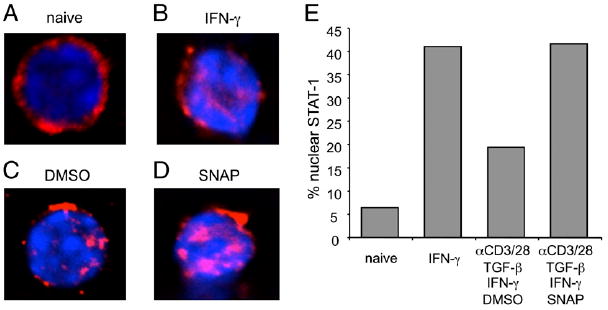
FIGURE 5.

NO augments IFN-γR signaling in Th cells. Naive OT-II CD4 T cells were treated with PBS (A) or as a control IFN-γ (B, 20 ng/ml). Other T cells were stimulated with anti-CD3/CD28, IL-2, TGF-β (1 ng/ml), and IFN-γ (1 ng/ml) in the presence of DMSO (C) and SNAP (D, 10 μM). Cells were fixed and stained with anti–STAT-1 (red) and DAPI (blue). Original magnification ×60. The percentages of cells having nuclear STAT-1 proteins were analyzed (E). Results are representative of three experiments.
