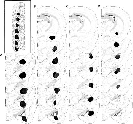
Figure 2.
Schematic representation of the minimum (gray) and maximum (black) extent of damage of unilateral excitotoxic lesions to the (A) posterior divisions of the basolateral and basomedial nuclei of the amygdala (BAp), (B) anterior divisions of the basolateral and basomedial nuclei of the amygdala (BAa), (C) lateral nucleus of the amygdala (LA), and (D) central nucleus of the amygdala (CE). The extent of unilateral electrolytic lesions of the amygdala is depicted in the inset. Brain images are adapted from Swanson (1999).

