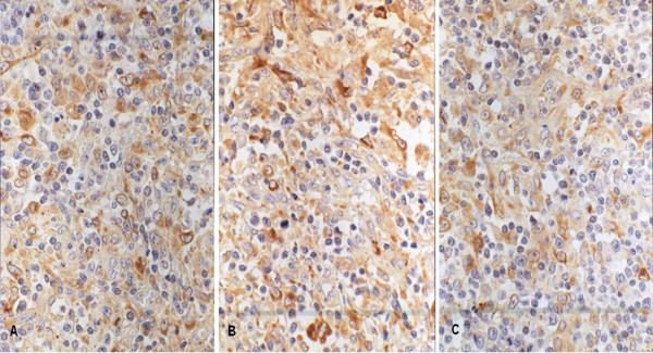
Figure 1.
The expression of CCR7, MMP-9 and MMP-2 in T-NHL with immunohistochemical staining. These markers all express in the cytoplasm. Some yellow or brown yellow granules in the cytoplasm are postive. The immunohistochemical staining was performed with S-P method and these photoes were taken under the high power (×400). A was CCR7 stainting. The staining intensity is strong. B was MMP-9 stainting. The staining intensity is strong. C was MMP-2 staining. The staining intensity is intermediate.

