
FIGURE 1:
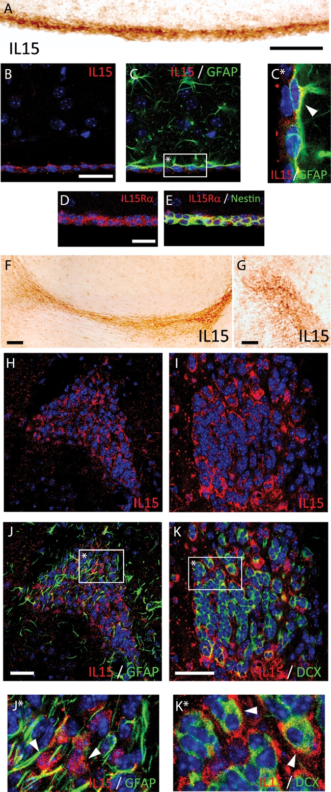
IL-15 expression in the SVZ and RMS. (A, F–G) Immunostaining of IL-15+ cells in the SVZ (A) and the RMS (F, sagittal; G; coronal). (B, C) Double immunofluorescence for IL-15 (red; B, C) and GFAP (green; C) in the SVZ. (C*) Inset: magnification of IL-15 colocalization in GFAP+ cells (white arrowhead). (D, E) Double immunofluorescence for IL-15Rα (red; D, E) and nestin (green; E) in the SVZ. (H–K) Double immunofluorescence for IL-15 (red; H, J and I, K) and GFAP (green; J) or DCX (green; K) in the RMS. (J*, K*) Inset: magnification of IL-15 colocalization in GFAP− cells (J*; white arrowhead) and DCX+ cells (K*; white arrowhead). Nuclei are stained with Hoechst (blue). Fluorescent sections are evaluated with confocal microscopy. Scale bar in A–E and G–K, 50 μm (shown in A, B, D, G, J, K); in F, 100 μm.