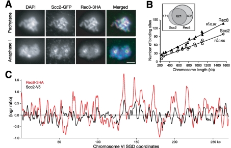
FIGURE 1:
Chromosome association of Scc2 and Rec8 during yeast meiosis. (A) Immunofluorescence of Scc2-GFP and Rec8'3HA (strain HY2020). Yeast cells were induced to undergo synchronous meiosis, and nuclear surface spreads were prepared and stained with GFP and HA antibodies. Two representative stages, pachytene and anaphase I, are shown. Red, Rec8'3HA; green, Scc2-GFP; blue, DAPI. Bar, 2 μm. (B) The number of chromosome-associated regions of Scc2 and Rec8 was proportional to chromosome length. Chromatin immunoprecipitation combined with microarray was used to identify Scc2 and Rec8 chromosomal binding during meiosis (strain HY1644). Inset shows the overlap of the Scc2 and Rec8 binding sites. (C) Visual representation of chromosome association profile of Scc2 and Rec8 during yeast meiosis. The entire chromosome VI is shown as a representative. The scale of the y-axis is log2 ratio of immunoprecipitation to input. SGD coordinates of chromosome VI are shown at the bottom. Red, Rec8 ChIP; black, Scc2 ChIP.

