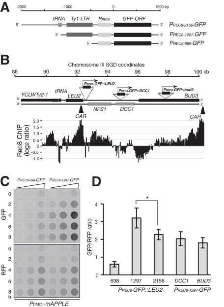
FIGURE 5:
Positional effect on REC8 promoter activation during meiosis. (A) A schematic diagram showing the three PREC8-GFP constructs. The only difference among them is the length of the 5′ upstream sequence. (B) Chromosomal location of the PREC8-GFP constructs. A schematic diagram showing the gene structure at the LEU2 locus on chromosome III. The location and orientation of the PREC8-GFP constructs are marked. Black bars show cohesin enrichment on the chromosome as determined by ChIP on chip. The scale of the y-axis is log2 ratio of immunoprecipitation to input. Two CARs in this region are depicted as black triangles. (C) GFP detected by a fluorescence scanner. Yeast cells were induced to undergo meiosis, and an aliquot was withdrawn at the indicated time, fixed in 1% formaldehyde, 1:2 serially diluted, and scanned with a fluorescence scanner. All strains harbored one copy of PDMC1-mAPPLE (RFP), which served as an internal control. Representative images are shown. (D) Quantitative analysis of PREC8-GFP gene expression. Three constructs were inserted at the LEU2 locus: PRec8'698-GFP (HY2666), PRec8'1297-GFP (HY2665), and PRec8'2158-GFP (HY2664) as shown in A. PREC8-GFP::DCC1 (HY2953) and PREC8-GFP::BUD3 (HY2952) were inserted at the DCC1 and BUD3 loci, respectively. GFP/RFP ratios were derived from samples at the 4-, 6-, and 8-h time points. Error bars show SD. *p value < 0.05, Student's t test.

