FIGURE 6:
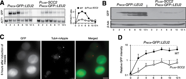
A reporter assay showing that Scc2 modulates REC8 promoter activity. (A) Meiotic expression of PREC8-GFP::LEU2 (HY2157 and HY2228). Yeast cells were induced to undergo meiosis, and samples were prepared for Northern blots as in Figure 3D. Quantification is shown to the right. (B) GFP protein level by immunoblot. Note that only minimal GFP protein can be detected in Scc2-depleted cells. (C, D) A quantitative-microscopy method of determining GFP level in live meiotic cells. Wild-type (HY2157) and PCLB2-SCC2 (HY2301) cells were mixed and induced to undergo meiosis, and 2-μl aliquots were withdrawn at indicated time points and mounted on a microscope slide for fluorescence microscopy. The PCLB2-SCC2 cells harbor one copy of γ-tubulin–mApple, which distinguishes them from the wild-type cells in the same microscopy field. Projected images from 14 Z-stacks are shown. Exposure for each section is 80 ms. Quantification of GFP intensity from each strain is shown in D. Error bars show SD. At least 50 cells were analyzed at each time point. Wild type, filled circles; PCLB2-SCC2, open circles. Bar, 4 μm.

