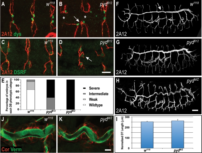
Figure 7:
Pyd is required for tracheal dorsal branch fusion and cell fate specification but not for formation or maintenance of adherens junctions. (A, B) Many dorsal branches (DBs) in zygotic pydB12 mutants fail to fuse (join to contralateral branch, asterisks in B), and often incorrectly fuse to neighboring branches (arrow in B). Anti-Dysfusion (Dys; green) labels fusion cells. (C, D) Labeling for the terminal cell marker DSRF reveals that some DBs in pydB12 embryos have more than one terminal cell (arrow in D), whereas control w1118 embryos have only one terminal cell per DB (C). (E) Quantification of DB defects in zygotic and maternal/zygotic (M/Z) pydB12 mutant embryos. DB fusion phenotypes are categorized into four classes as in Jung et al. (2006): wild type (complete dorsal branch fusion), weak (only one affected metamere), intermediate (two to four affected metameres), and severe (five or more affected metameres). (F–I) Dorsal trunks (DTs, arrow in F) of zygotic and M/Z pydB12 mutants are contiguous, indicating normal adherens junction function and fusion of dorsal trunk segments. (H–I) DTs of pydMZ embryos appear tortuous but are the same length as w1118 control embryos (p = 0.20, Student's t test). DT length is normalized to embryo length. (J, K) pydMZ mutants have normal localization of the septate junction marker Coracle (Cor) and normal lumenal accumulation of the matrix marker Verm. Scale bars, A–D, 5 μm; F–H, 20 μm; J, K, 10 μm.

