Figure 9:
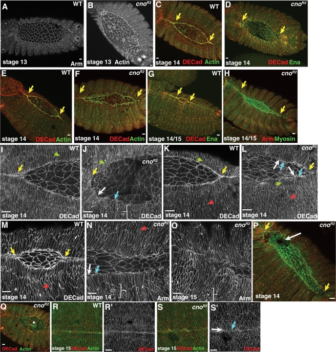
cno zygotic null mutants have defects in dorsal closure and cell shape change that are very similar to those of pydMZ mutants. Stage 13–15 wild-type (WT) embryos or cnoR2 zygotic mutants, anterior left, dorsal view, antigens, genotypes and stages indicated. (A, B) Onset of dorsal closure. cno mutants appear quite normal. (C–F) Early stage 14. cno mutants have an uneven leading edge and reduced zippering at the canthi (yellow arrows). (G, H) Those defects continue as closure moves to completion. Note the extreme retardation of zippering at canthi in mutants (yellow arrows). (I–O) Close-ups, stage 13 (I, J), early stage 14 (K, L), or late stage 14 (M–O) embryos. Whereas LE cells (green arrows) and more lateral epidermal cells (red arrows) can elongate, substantial defects are seen in cell shape. LE cells often have broadened (blue arrows) or hyperconstricted (white arrows) leading edges, some cells fail to change shape (brackets), and the leading edge of the epidermal sheets is very wavy. (P, Q) In some embryos, the amnioserosa ruptures before completion of dorsal closure (P, white arrow; Q, asterisk). Yellow arrows indicate retarded zippering. (R, S) stage 15. In the subset of mutants completing closure, cell shapes remain irregular (blue arrow) and small dorsal discontinuities are seen (white arrow). Scale bars, 10 μm.

