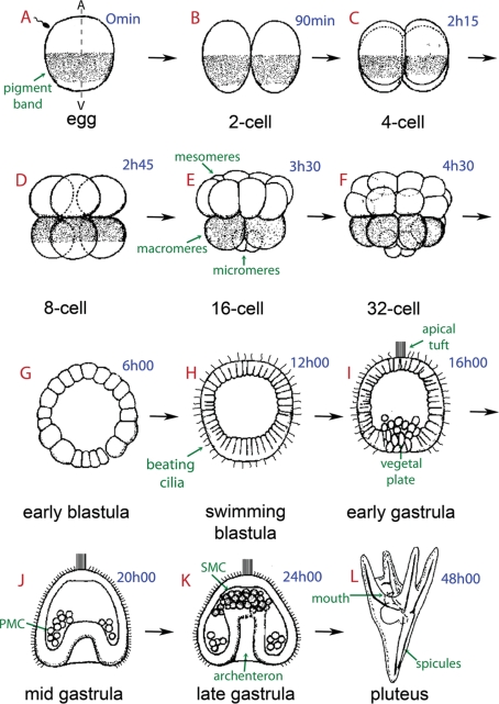
Figure 1:
Schematic representation of sea urchin embryonic development. The times in blue indicate hours postfertilization. The sea urchin oocyte is organized along an animal–vegetal (A/V) primary axis, which is observable in P. lividus due to the presence of a subequatorial cortical pigmented band visible under blue light (arrow in A). Fertilization is followed by first and second cleavages, which are meridional (parallel to the A/V axis) and perpendicular to each other (B, C). The third cleavage is equatorial, perpendicular to the first two cleavage planes, and separates the animal and vegetal hemispheres from one another (D). During the fourth cleavage, animal blastomeres divide equally to produce eight mesomeres, and the vegetal blastomeres divide unequally to produce large macromeres and small micromeres located at the vegetal pole of the embryo (E, F). Six hours after fertilization the sea urchin embryo enters the early blastula stage with an empty central cavity called a blastocoel (G). The cells start developing cilia on their outer surface to form a swimming blastula (H). Approximately 10–12 h after fertilization, the midblastula, composed of ∼600 cells, hatches out of the fertilization envelope (not shown). At the animal pole, the cilia are longer but do not beat. This “apical tuft” (I) provides directionality to swimming, as embryos almost always move with the apical tuft region forward. In the late blastula stage the embryo becomes thickened at the vegetal pole, forming the vegetal plate (I). This represents the gastrulation site where the primary mesenchyme cells (PMCs), which are derived from the micromeres and located in the center of the vegetal plate region, migrate into the blastocoel (I, J). The vegetal cells will continue to ingress in order to form the archenteron, led by filopodia extending from the secondary mesenchyme cells (SMCs), which eventually contact the animal pole at the future site of mouth formation (K). A prism (not shown) and finally a feeding pluteic larva will be formed 24 h later around an endoskeleton, which contains two spicules made of calcium carbonate secreted by the PMCs (L). A few days later, this pluteus will metamorphose into a tiny male or female adult urchin.

