Figure 2:
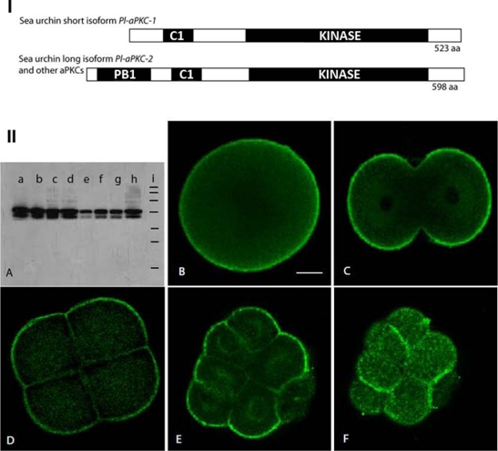
I. Domain structure of sea urchin aPKCs. (A) The short isoform found so far in P. lividus and S. purpuratus encodes an aPKC that lacks the PB1 protein interaction domain. Kinase: serine threonine kinase domain; C1: cysteine-rich domain, which binds InsPtd(3,4,5)P3. The three urchin species examined so far (P. lividus, H. pulcherrimus, and S. purpuratus) also contain a long isoform that is similar to most other known aPKCs in its domain composition (see Supplemental Figure S1 for alignment). II. Two maternal aPKC proteins that localize in the cortex in early embryos of P. lividus. (A) Western blot with the SC216 anti-aPKC antibody on P. lividus extracts from (a) unfertilized eggs, (b) 2-cell stage, (c) 8-cell stage, (d) 16- to 32-cell stage, (e) swimming blastula, (f) early gastrula, (g) prism, (h) plutei. Equal amounts of protein were loaded in each lane. (i) Standard molecular weight markers from top to bottom: 200, 120, 100, 70, 50, 37, and 20 kDa. (B–F) Immunolocalization of aPKC in early sea urchin embryos observed by confocal microscopy. (B) Unfertilized egg, (C) 2-cell stage, (D) 8-cell stage, (E, F) two confocal sections of the same 16-cell embryo showing (E) the interior and (F) the surface. Scale bar, 20 μm.

