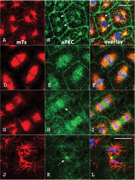
Figure 3:
aPKC is associated with microtubule structures during mitotic divisions in the early sea urchin embryo. Sixty-four–cell stage embryos were fixed and labeled with DM1A antitubulin antibody (red) and anti-aPKC antibody (green). aPKC associates with the MTOC (arrows in B and C) during prophase (A–C); decorates spindle microtubules during metaphase (D–F); starts to concentrate in the spindle midzone (arrow) during anaphase (G–I), and ends up in the midbody (arrow) during telophase (J–L). The nucleus (in C) and chromosomes (in F, I, and L) are stained in blue with Hoescht stain. Bar, 5 μm.

