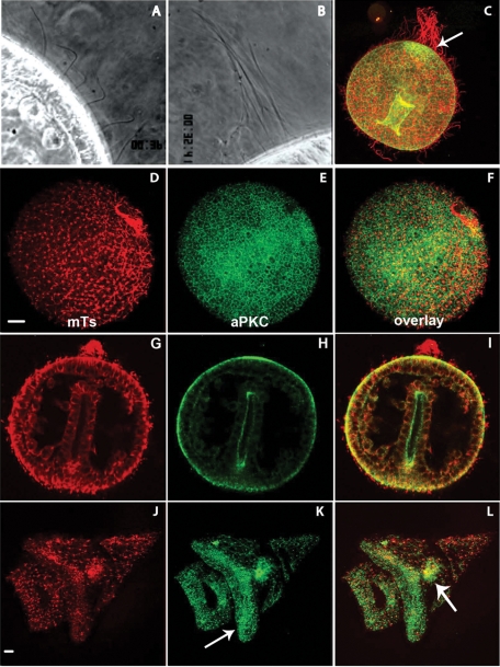
Figure 4:
aPKC localization in ciliated swimming sea urchin embryos. (A, B) Dark-field videomicroscopy showing (A) short, active cilia and (B) the “apical tuft” made of a few long and immobile cilia at the animal pole. (C–L) Confocal sections of swimming embryos fixed and labeled for microtubules (red) and aPKC (green). (C) Early gastrula showing the longer cilia (in red) and the concentration of aPKC at the animal pole (arrow). (D–F) Swimming blastula. (G–I) Late gastrula. (J–L) Pluteus larva; arrows: ciliary band in K and stomach in L.

