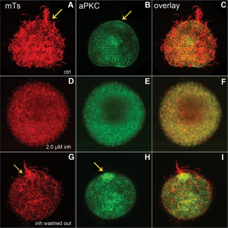
Figure 8:
The inhibition of aPKC and its effect on ciliogenesis can be reversed. (A–C) Control embryos were deciliated and left to reciliate for 2 h before fixation and staining for microtubules (red) and aPKC (green). The arrows show the apical tuft and the accumulation of aPKC at the animal pole. (D–F) Embryo deciliated and left to reciliate in the presence of 2.0 μM pseudosubstrate, fixed, and stained like the control. (G–I) Embryo treated as in D, then washed to remove the inhibitor and allowed to reciliate for 2 h. The arrows show the reformation of the apical tuft and apical accumulation of aPKC when the kinase is no longer inhibited.

