FIGURE 1:
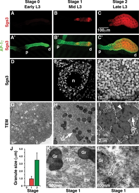
Glue granule biogenesis is developmentally regulated. (A–C′) Confocal micrographs of whole third-instar larval (L3) salivary glands expressing Sgs3-DsRed (red) and stained for AP-1γ (green), showing developmental timing of Sgs3-DsRed expression from stage 0 (no granules) through stage 1 (initiation of granule production) to stage 2 (fully mature granules or glands). AP-1γ is expressed in all cells of the salivary gland throughout development, whereas Sgs3-DsRed is first detected in distal (d) mid-L3 salivary gland cells (B, B′) and is expressed in more proximal (p) cells as development proceeds (C, C′). (D–F) Confocal micrographs of individual salivary gland cells showing developmental expression of Sgs3-DsRed. Sgs3-DsRed is not expressed in stage 0 (D). In stage 1, granules surround the nucleus (n) and appear uniformly small (E). In stage 2, granules are larger and occupy most of the cytoplasmic space (F). (G–I) Transmission electron micrographs (TEM) of L3 salivary glands staged using the Sgs3-DsRed marker. No granules were detected in stage 0 (G). Glue granule (Gr) maturation observed by TEM (H, I) parallels that seen by Sgs3-DsRed, validating this marker for following glue granule biogenesis (E, F). (J) Granules increase in size over time, from an average length of 1.0 μm ± 0.3 (n = 91) at stage 1 (red bar) to a maximum length of 3.5 μm ± 1.0 (n = 54) at stage 2 (green bar). (K, L) TEM of stage 1 salivary gland cells. Rough ER, transitional ER (tER), Golgi, and TGN (defined morphologically as in the work of Thomopoulos et al., 1992; Kondylis and Rabouille, 2009) are present near small glue granules (Gr) (K). Coated vesicles (CV) were also observed near glue granules (Gr) (L).

