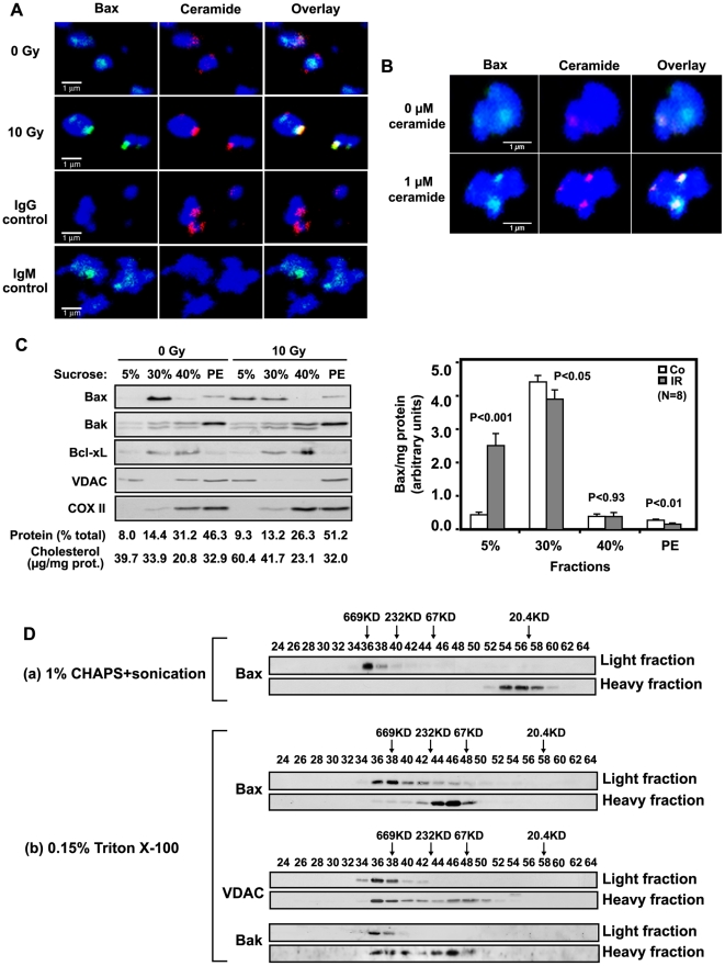
Figure 5. Ceramide induces formation of a mitochondrial ceramide-rich macrodomain (MCRM).
(A) Ionizing radiation (10 Gy) induces co-localization of endogenous Bax with MCRMs in HeLa cells. Mitochondria were isolated from HeLa cells 34 h after irradiation and immunostained as described in Supporting Information Text S1. Data represent typical stainings from 1 of 4 similar studies in which 2000 mitochondria were analyzed each. (B) Addition of exogenous C16-ceramide induces co-localization of endogenous full-length Bax with MCRMs in HeLa cells. Mitochondria were isolated from HeLa cells using percoll gradient and treated with ceramide as Figure 3A. After 30 min incubation, mitochondria were fixed and stained with MitoTracker (blue), while ceramide and Bax were localized using anti-ceramide IgM (red) or anti-Bax IgG (green), respectively. Control IgM and IgG did not yield detectable signals (not shown). These data represent 1 of 3 similar studies. (C) Bax translocates into a radiation-generated HeLa MCRM. Upper panel: 34 h post-irradiation, HeLa mitochondria were isolated as in Materials and Methods and incubated with 0.15% Triton X-100 in MBS buffer for 30 min on ice. 40 µl mitochondrial homogenate (3.3 µg/µl) were subjected to 5–30% mini-discontinuous sucrose density gradient centrifugation as described in Materials and Methods. 20 µl aliquots of 80 µl fractions were analyzed by immunoblotting using the indicated antibodies. The protein level of each fraction was assessed using the Bio-Rad Dc protein assay kit (PE, Pellet). Data are from 1 of 4 studies, consisting of 2 independent gradients per study. The gradient shown displays our clearest example of Bax translocation into light membranes. Lower panel: Bax in each fraction, revealed by immunoblotting and quantified using NIH Image software, was normalized to protein content for all 8 gradients. (D) MCRM Bax exists as high molecular weight oligomers. Mitochondria from 10 Gy-irradiated HeLa cells, disrupted by either (a) 1% CHAPS and sonication or (b) dounce homogenization in 0.15% Triton X-100, were subjected to 5–30% discontinuous sucrose gradient for MCRM isolation as in Experimental Procedures. Light (MCRM; fractions 6,7) and heavy fractions (solubilized proteins; fractions 11,12) were analyzed by gel filtration on Sephacryl S-200 column as in Figure 2C. 500 µl of each eluted fraction were concentrated by 20% TCA precipitation for immunoblotting. Data are from 3 independent studies.

