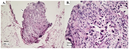
Figure 1. Haematoxylin and eosin staining shows extranodal metastasis (EM) in gastric carcinoma.
Tumor cells are scattered into the resected adipose connective tissue around the stomach distinct from the metastatic lymph node. 1A: Original magnification ×100, arrow indicates the EM. 1B: Original magnification×400.

