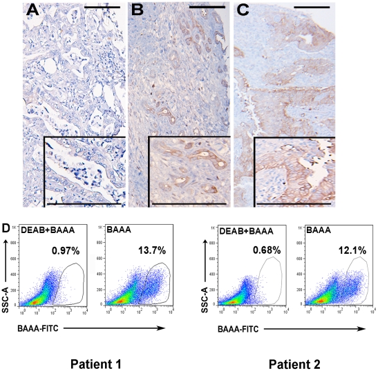
Figure 2. ALDH1 expression and activity in untreated primary pancreatic cancer specimens.
Representative images of heterogeneous ALDH1 expression among patients with pancreatic cancer. Analysis of a TMA comprised of 106 untreated pancreatic tumors demonstrated (A) low ALDH1 expression in 22% (23/106) of examined patient specimens. (B) Moderate ALDH1 expression was detected in 26% (28/106) of examined patient specimens. (C) High ALDH1 expression was detected in 22% (23/106) of examined patient specimens. (D) Flow cytometry results of patient pancreatic adenocarcinoma tumors after digestion into a single cell suspension and stained with Aldefluor reagent, with and without DEAB inhibitor. Populations of cells with high ALDH activity relative to the overall cell population are easily distinguished. ALDHhigh cells = 12.7% (Patient 1) and 11.4% (Patient 2) of all viable human cells. Scale bar = 250 µm.

