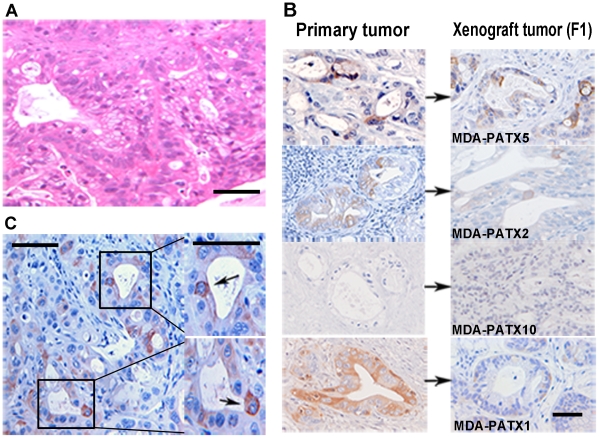
Figure 3. Histologic analysis of patient and direct xenograft tumors for expression of ALDH1.
(A) Representative image demonstrating the histologic appearance of direct xenograft tumors established from freshly resected pancreatic tumors. Note tumor-gland formation and associated peri-tumoral stroma. (B) Comparison of ALDH1 expression in four different direct xenograft tumors to ALDH1 expression in original (parental) patient tumors. The pattern and location of ALDH1 expression is maintained during the xeno-transplantation process as reflected in derived xenograft tumors. An example of undetectable ALDH1 expression in both the patient tumor and derived direct xenograft is shown in the third panel from the top (MDA-PATX10). (C) Intra-tumoral heterogeneity of ALDH1 expression in direct xenograft tumors is readily identified as only a subset of luminal tumor cells demonstrate intense staining for ALDH1 relative to all other cells within tumor. Scale bar = 125 µm (A), 180 µm (B), 125 µm (C).

