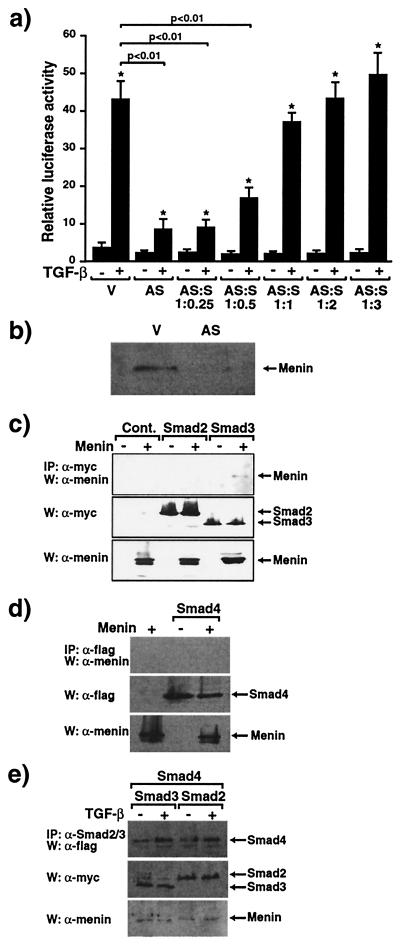
Figure 3.
Antisense menin inhibits TGF-β-mediated transcriptional responses and menin specifically binds Smad3. (a) 3TP-Lux was transfected into HepG2 cells together with empty vector (V) or antisense menin (AS) either alone or with sense menin (S) and the cells were stimulated (+) or not (−) with 200 pM TGF-β. Relative luciferase activity was measured. Values of relative luciferase activity represent the mean ± standard error of three separate experiments. *, P < 0.01, TGF-β + versus − and, shown as a bar, P < 0.01, AS versus V. (b) Menin expression in empty vector (V)- and antisense menin (AS)-transfected HepG2 cells assessed by immunoblotting. (c–e) Association of menin and Smad proteins. W, Western blot; IP, immunoprecipitation. Menin was transfected into COS7 cells with the indicated myc-tagged Smad constructs (c and e) or flag-tagged Smad4 (d and e). Cell extracts were immunoprecipitated with anti-myc (c), anti-flag (d), or anti-Smad2/3 antibodies (e) followed by immunoblotting with menin (c–e) or anti-flag (d) antibodies. Expression of myc, flag, or menin was monitored.

