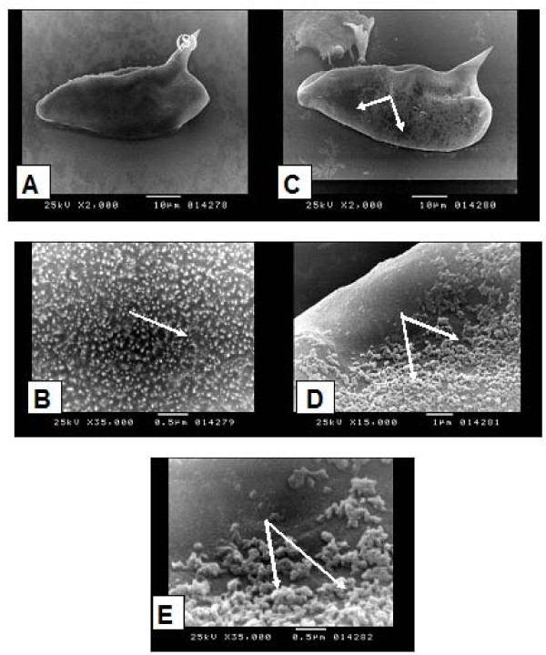
Figure 2.
Scanning electron microscopy (SEM) of Schistosoma mansoni eggs: unexposed eggs showing: (A) oval egg with lateral spine (S) (× 2000), (B) the surface of the egg showed microspicules like chitinous projections (→) (× 35000). Exposed eggs to miltefosine showing: (C) patchy loss of the microspicules like chitinous projections (→) (× 2000), (D) higher magnification of (C) (× 15000), (E) swollen and oedematous chitinous projections (→) (× 35000).

