
Figure 7.
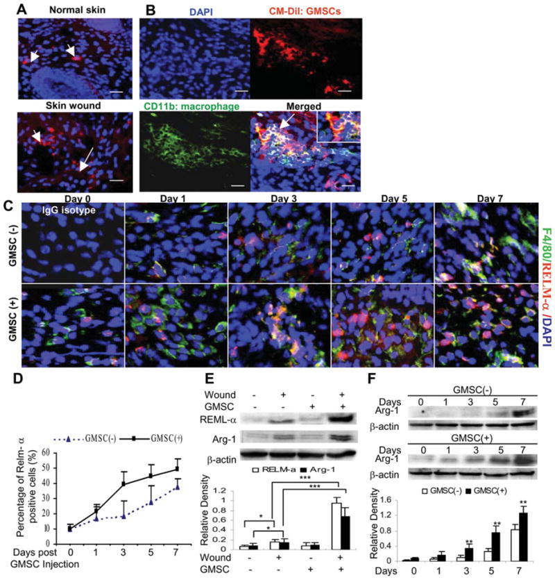
Interactions of homed GMSCs with macrophages during wound healing. (A): GMSCs prelabeled with CM-DiI were systemically infused by tail vein (i.v.) into mice one day after skin wounding. Seven days after cell injection, skin tissues were frozen sectioned and observed under a fluorescence microscope, whereby normal skin on the other side of the same mice were used as controls. (B): Frozen sections of wounded skins from mice after injection with CM-DiI pre-labeled GMSCs were immunostained with fluorescein isothiocyanate-conjugated antibody for mice CD11b. Scale bar = 50 μm. The results were representative of at least three independent experiments. (C): Frozen sections of full-thickness incisional skin wounds from mice after treatment with GMSCs for different days were dual-color immunostained with specific antibodies for F4/80 (Green) and RELM-α (Red). Scale bar = 50 μm. (D): Quantification of M2 macrophages positive for RELM-α. (E): Western blot analysis of arginase-1 (Arg-1) and RELM-α expression in skin wounds 7 days post GMSC treatment. (F): Time-dependent increases in Arg-1 and RELM-α expression induced by GMSC treatment. The results are representative of three independent experiments. *, p < .05; **, p < .01; ***, p < .001. Abbreviations: DAPI, 4′, 6-diamidino-2-phenylindole; GMSC, mesenchymal stem cells from human gingival; RELM, resistin-like molecule.