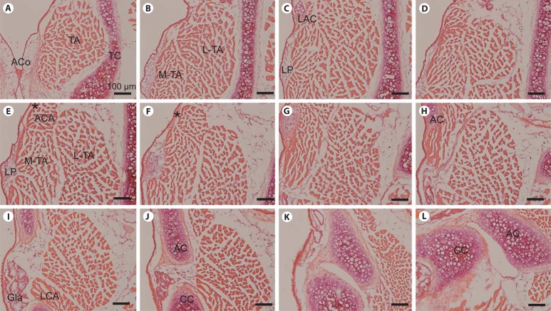
Fig. 1.
Representative H&E-stained images of serial whole-mount coronal sections from a wild-type FVB control mouse larynx. A–L Panels are ordered from anterior to posterior; the distance between each panel is approximately 120 μm. AC = Arytenoid cartilage; ACA = alar cricoarytenoid muscle; ACo = anterior commissure; CC = cricoid cartilage; Gla = subglottal gland; LAC = laryngeal alar cartilage; LCA = lateral cricoarytenoid muscle; LP = lamina propria; L-TA = lateral thyroarytenoid muscle; MTA = medial thyroarytenoid muscle; TA = thyroarytenoid muscle; TC = thyroid cartilage. Asterisk indicates ligament-like structure extending from the LAC to the AC. Scale bar: 100 μm.

