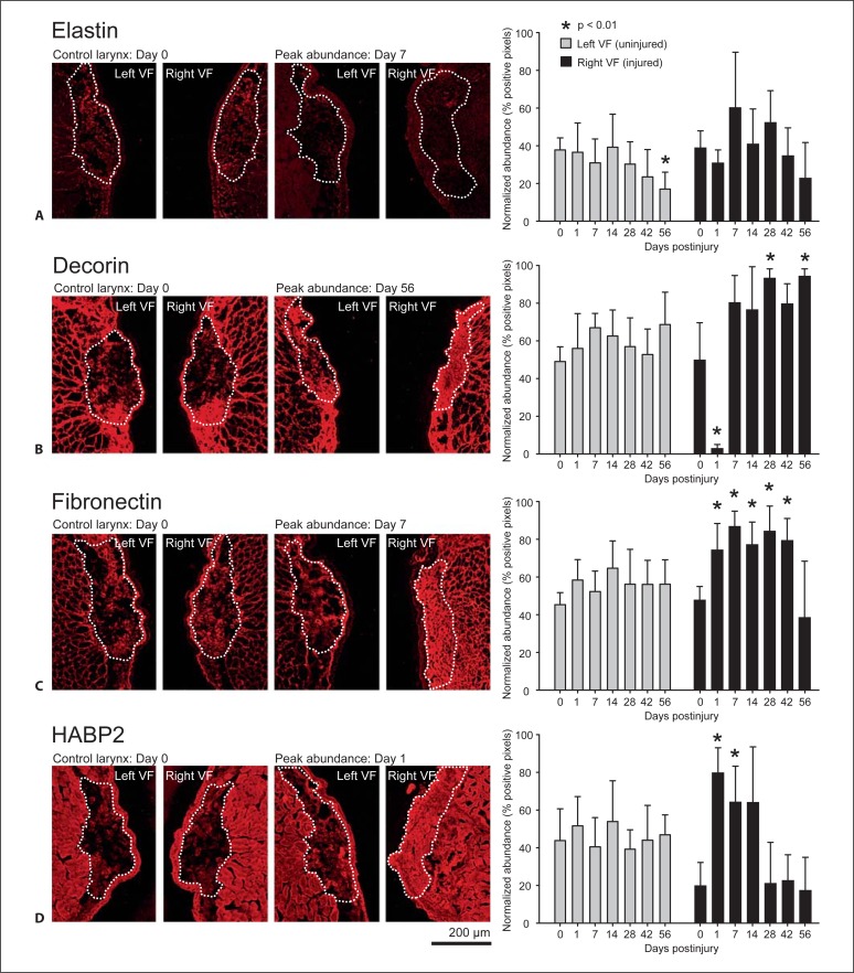
Fig. 3.
Representative IHC images and quantitative analysis of elastin (A), decorin (B), fibronectin (C), and HABP2 (D) in uninjured control and injured vocal fold LP over time. Normalized abundance is expressed with respect to total LP area (indicated by a white dashed line). Error bars represent standard deviations and asterisks denote statistically significant differences (p < 0.01) from the control group (day 0). VF = Vocal fold. Scale bar: 200 μm.

