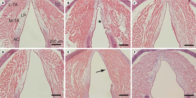
Fig. 4.
Representative H&E-stained images of whole-mount axial sections from an uninjured control mouse larynx and unilaterally injured mice larynges at 1, 7, 14, 28 and 42 days postinjury. A Uninjured control. B One day postinjury. C Seven days postinjury. D Fourteen days postinjury. E Twenty-eight days postinjury. Arrow indicates dense eosinophilic region and LP contraction. F Forty-two days postinjury. H&E-stained whole-mount coronal sections from additional mice at each time point revealed comparable histopathology (data not shown). AC = Arytenoid cartilage; LP = lamina propria; L-TA = lateral thyroarytenoid muscle; M-TA = medial thyroarytenoid muscle; TC = thyroid cartilage. Asterisk indicates fibrin clot and infiltration of inflammatory cells. Scale bar: 200 μm.

