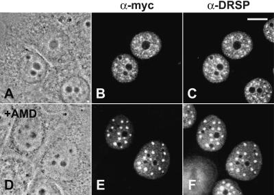
Figure 3.
Intracellular localization of myc-tagged wt SF3b155 from X. laevis transiently expressed in human MCF-7 cells in the absence (A–C) and presence (D–F) of actinomycin D. Phase contrast micrographs (A and D), immunofluorescence micrographs upon incubation with the anti-myc antibody (B and E), and the antibody directed against the DRS protein (23), respectively (C and F). Scale bar is 10 μm.

