
Figure 3.
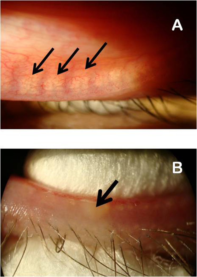
Visible light microscopy evaluation of meibomian glands (panel A) and manual expression of meibomian gland secretions from a lower eyelid of a volunteer using two cotton swabs.
Panel A. Meibomian glands are marked with black arrows.
Panel B. Expressed meibum is marked with a black arrow.