
Figure 5.
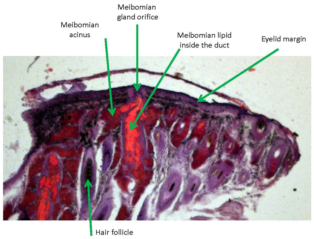
Histochemical staining of meibomian glands. An upper mouse meibomian gland is shown. The tissue lipids were stained with Oil Red O and counter-stained with hematoxylin. Notice accumulation of large amount of stained lipids (bright red) in the main (central) duct of the gland.