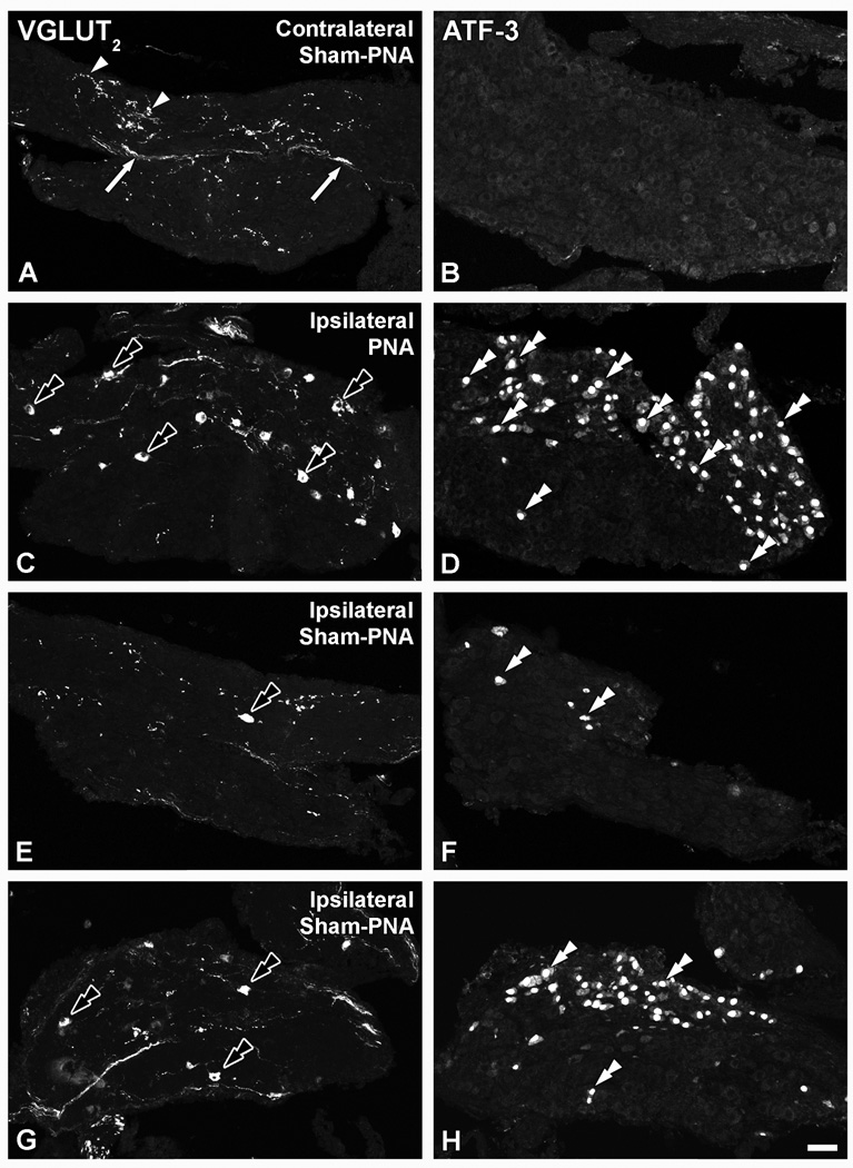
Figure 3.
Confocal immunofluorescence photomicrographs of sections of the contralateral (A, B) or ipsilateral (C–H) LSC of mice 7 days after sham-PNA (A,B; E–H) or PNA (C, D), incubated with VGLUT2 (A, C, E, G) or ATF-3 (B, D, F, H) antisera (each corresponding pair of VGLUT2 or ATF-3 micrographs shows sections separated by 36 µm). Arrows and arrowheads in (A, B) show VGLUT2-IR fibers and varicosities, respectively. Black double arrowheads and double arrowheads in (C, D, E–H) show VGLUT2- and ATF-3-IR LSC NPs. Scale bar: 50 µm (H=A–G).

