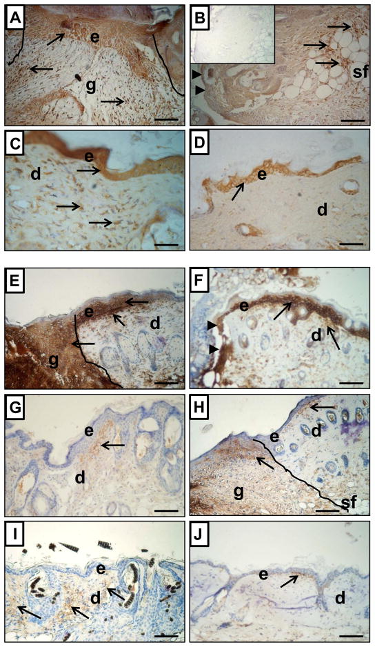
Figure 6.
Immunohistological detection of MMP-9 (A–D) and MMP-13 (E–J) expression in skin samples from Hsd: Athymic Nude-Foxn1nu (A, C, E, G and I) and wild-type BALB/c (B, D, F, H and J) mice at post-wounded Day 3 (A, B, E and F), Day 7 (G–H) and Day 36 (C–D and I–J). The post-wounded area is outlined on A, E and H. Arrowheads indicate the margins of opened/unhealed wound edges on B and F. Arrows indicate MMP-9 or MMP-13 positivity (brown deposits). Sections were counterstained with hematoxylin. Abbreviations: e, epidermis; d, dermis; sf, subcutaneous fat; g, granulation tissue. Insert in B shows control of immunoreaction where first antibody were replaced by non-specific IgG. Scale Bars: 100μm.

