
Figure 5.
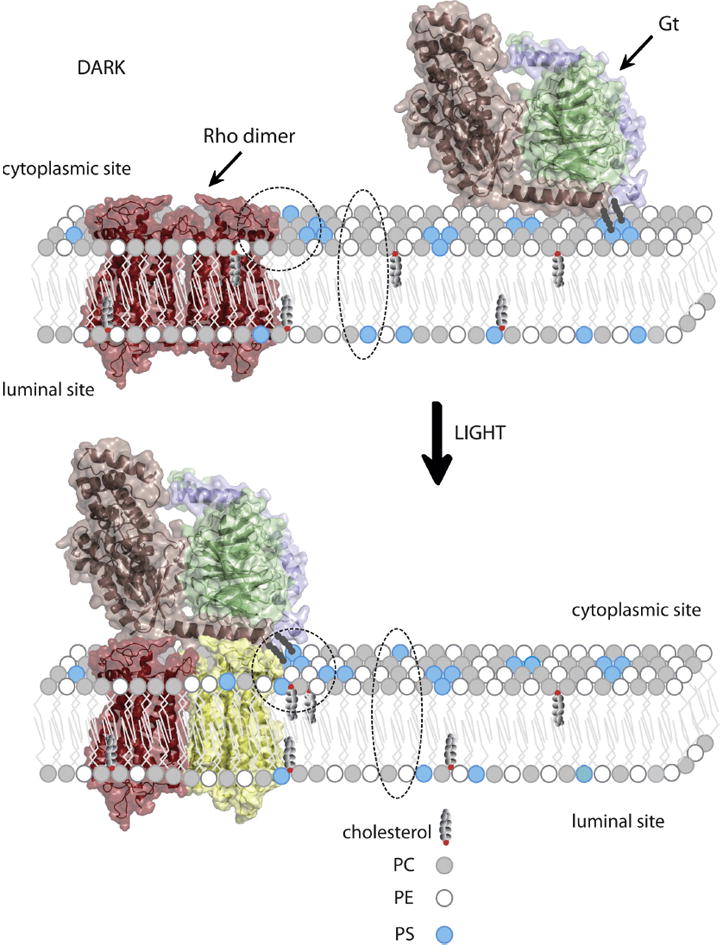
Reorganization of specific lipids in a disk membrane upon light illumination allows appropriate binding of Gt to photoexcited rhodopsin. Model of rhodopsin dimer (26, 39) is colored red in the dark state. Activated rhodopsin molecule is colored yellow. Gtα, Gtβ and Gtγ within heterotrimeric G protein are colored light pink, green and violet, respectively. Gt is attached to the membrane through myristoyl and farnesyl groups. Headgroups of phosphatidylcholine (PC), phosphatidylethanolamine (PE) and phosphatidylserine (PS) are depicted as small circles colored grey, white and blue, respectively. Movement of PS from the inner (lumenal) to the outer (cytoplasmic) leaflet of the lipid bilayer, as well as reorganization of PS, PE and cholesterol in close proximity to the activated rhodopsin molecule, is highlighted with a broken ellipsoid and circle, respectively. This lipid reorganization plays an important role in formation of the rhodopsin-Gt complex, thereby activating the visual signaling cascade.