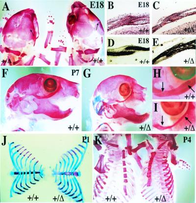
Figure 3.
Precocious ossification of coronal sutures and the sternum in FgfR2-IIIc+/Δ mice. Skeletons stained with alizarin red to identify ossified tissue (A and K), or stained with alizarin red in combination with Alcian blue stain to additionally reveal cartilage (F–J). (A) Dorsal view of calvarial bones at E18 showing closer apposition of frontal and parietal bones at the coronal suture (arrows) in a hemizygote compared with its wild-type litter mate. (B–E) Transverse sections through the frontal and parietal bones showing fusion of coronal sutures in hemizygote in contrast to wild-type mice. Sections stained with hematoxylin–eosin (B and C) or alkaline phosphatase (D and E). (F–I) Lateral views of skulls from 7-day-old mice showing in hemizygotes rounded heads, truncated maxilla, and fusion of joints separating the zygomatic arch bones (part of the maxilla, zygomatic, and temporal bones) which make up the lower rim of the eye socket. (H and I) Detail of zygomatic arch joints shown in F and G, respectively. (J and K) Dissected and whole rib cages from 1- and 4-day-old mice, respectively, showing precocious and progressive sternal fusion in hemizygotes. Note that in these mice, individual sternebrae are thicker and less congruent, and the manubrium and xiphoid processes are bifurcated.

