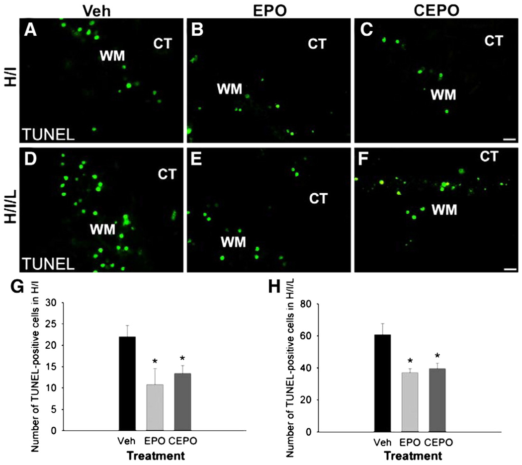
Fig. 7.
EPO and CPEO decrease white matter apoptosis after neonatal cerebral ischemia. Mice underwent H/I or H/I/L and received i.p. injections of EPO, CEPO or vehicle immediately after the induction of injury. We evaluated TUNEL immunoreactivity 24 h after treatment. TUNEL+ apoptotic cells were identified and counted in the white matter area. In both the H/I model (A–C) and the H/I/L model (D–F), EPO and CEPO reduced the appearance of TUNEL-positive cells. (G, H) We counted the TUNEL-positive cells and confirmed their number was significantly reduced in EPO or CEPO treatment groups compared with control groups (*, p<0.05). Scale bar, 10 µm. WM, white matter. CT, cortex.

