Fig. 8.

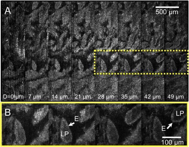
Images of a swine small intestine tissue: A. SECM images obtained from different depth levels; and B. magnified view of the dotted box in (A). LP – lamina propria; E – epithelium.

Images of a swine small intestine tissue: A. SECM images obtained from different depth levels; and B. magnified view of the dotted box in (A). LP – lamina propria; E – epithelium.