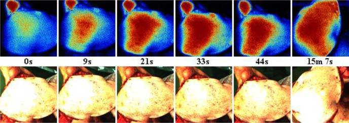
Fig. 5.

Reperfusion of a free flap of ~40 cm2 area. The image center corresponds to the perforator location, where arterial blood flow was reinitiated at time point zero. Within ~30s the microcirculation in the central region was restored. A final check after 15 min showed that the entire flap was sufficiently well perfused for the final flap insertion. The least-perfused region in the bottom-center of the perfusion maps corresponds to the position of a staple holding the tissue in place.
