Fig. 3.

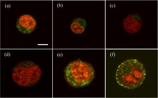
Typical confocal image slices acquired from cells with double fluorescent stains for nucleus and mitochondria: (a) Jurkat; (b) NALM-6; (c) U937; (d) MCF-7; (e) B16F10; (f) TRAMP-C1. Bar = 5μm.

Typical confocal image slices acquired from cells with double fluorescent stains for nucleus and mitochondria: (a) Jurkat; (b) NALM-6; (c) U937; (d) MCF-7; (e) B16F10; (f) TRAMP-C1. Bar = 5μm.