Figure 2.
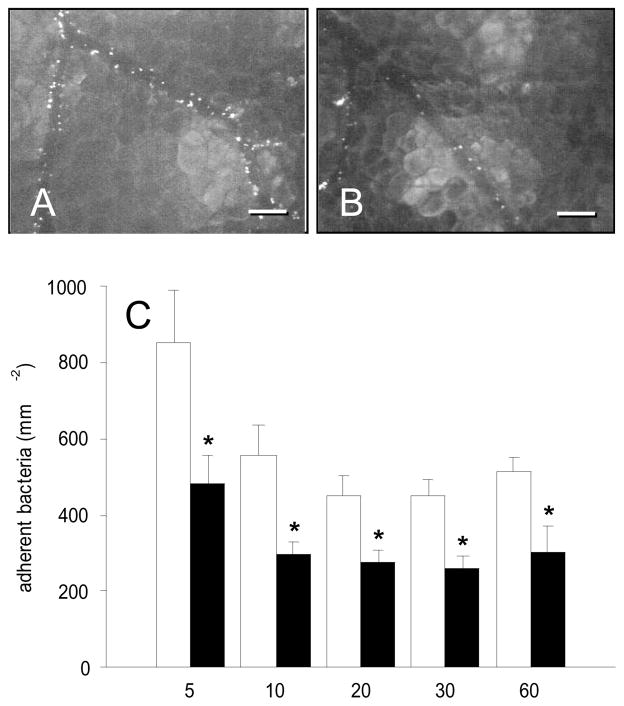
A, B: Intravital fluorescence microscopy of FITC-labeled Staphylococcus aureus in postcapillary and collecting venules of the dorsal skinfold chamber (5 min after intra-arterial injection of bacteria) after local TNF-α-exposure. Note that blockade of gC1qR by application of MAb 74.5.2 (B) results in a significant reduction of bacterial adherence when compared to controls (A). Blue light epi-illumination; scale bars: 100μm. C: Adherent bacteria (mm−2) in venules of TNF-α-exposed dorsal skin-fold chambers of Syrian golden hamsters, which were treated with MAb 74.5.2 (black bars; n = 7) or vehicle (control, white bars; n = 7), as assessed by intravital fluorescence microscopy and computer-assisted image analysis. Measurements were performed at 5, 10, 20, 30, and 60 min after intra-arterial injection of the bacteria. Values are given as means ± SEM; *p<0.05 vs. control.

