Figure 1.
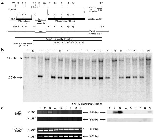
Generation of V1b receptor–deficient mice. (a) Simplified restriction map of the V1b receptor gene and structure of the targeting vector. The coding region of the exon is boxed. Neo, neo cassette; DT-A, diphtheria toxin A fragment gene; B, BamHI; E, EcoRI; EV, EcoRV; H, HindIII; S, SacI; Sp, SpeI; X, XhoI. (b) Southern blot analysis of tail DNA. DNA was digested with EcoRV and the blot was hybridized with the 5′ probe shown in a. The 14.0-kb band is derived from the WT allele (Wild) and the 2.8-kb band from the targeted allele (Mutant). (c) RT-PCR analysis of the RNA from tissues of V1bR+/+ and V1bR –/– mice. Ethidium bromide stainings of RT-PCR fragments are shown at left. The V1b receptor mRNA transcripts were detected and are shown as 540-bp fragments. The control for RT-PCR analysis was provided by detection of the 662-bp fragment of the GAPDH message. Southern blots of the RT-PCR fragments are shown at right. The specificities of the amplified fragments were assessed using 32P-labeled probes specific for each receptor subtype. Lane 1, brain; lane 2, hippocampus; lane 3, pituitary; lane 4, heart; lane 5, lung; lane 6, liver; lane 7, kidney; lane 8, spleen; lane 9, aorta.

