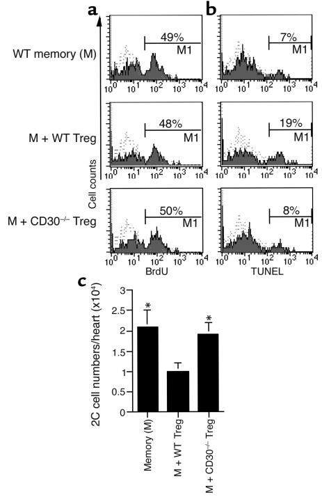
Figure 4.
Treg cells promote the apoptosis of memory CD8+ T cells but do not inhibit their proliferation in vivo. (a) Analysis of in vivo memory T cell proliferation by BrdU labeling. Splenectomized aly mice, transplanted with a cardiac allograft, received 1B2+CD8+ memory T cells and/or Treg cells and were pulsed intraperitoneally with BrdU 6 days after cell transfer. Twenty-four hours later, graft-infiltrating cells were stained for 1B2, CD8, and BrdU. The percentage of BrdU+ cells is shown in the histograms after gating on 1B2+CD8+ cells. The dotted lines represent isotype control. One of three experiments is shown. (b) Analysis of in vivo memory T cell apoptosis by the TUNEL method. Graft-infiltrating cells from the mice similar to those described above were stained for 1B2, CD8, and TUNEL. The percentage of TUNEL+ cells is shown in histograms after gating on 1B2+CD8+ cells. The dotted lines represent negative control. One of three experiments is shown. (c) Absolute number of memory 1B2+CD8+ T cells per graft was calculated according to flow cytometry. One of three experiments is shown. *P < 0.05 vs. the M + WT Treg group.

