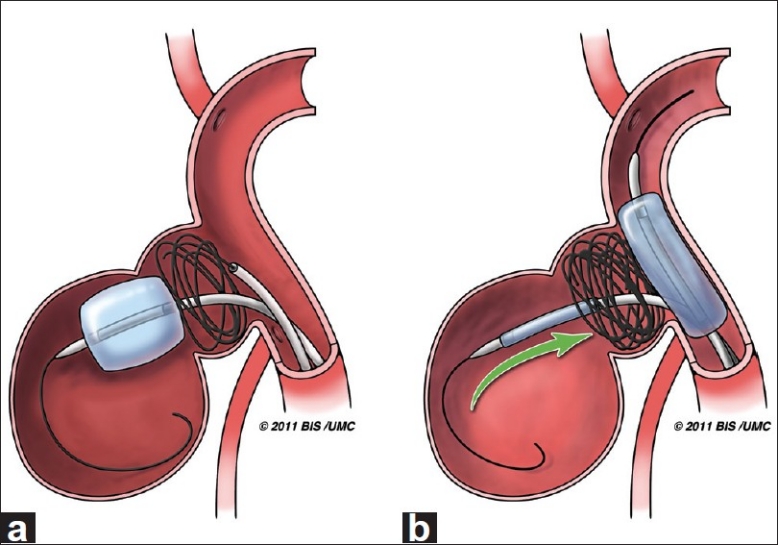
Figure 3.

Artist illustration of the intra-aneurysmal balloon-assisted coiling technique. (a) Inflated Hyperform balloon at the entrance of the larger distal aneurysmal lobule, limiting coiling to the proximal lobule. (b) A Hyperglide balloon is inflated across the aneurysm neck as the deflated intra-aneurysmal balloon is slowly removed to prevent coil mass disturbance (Artist: W. Kyle Cunningham, Medical Illustrator at the University of Mississippi Medical Center)
